

Ann Arbor-Based Dauer Lab Uses Mouse Models in Brain Disease Research
Dr. William Dauer’s busy lab at the University of Michigan pursues cutting-edge neuroscience research.
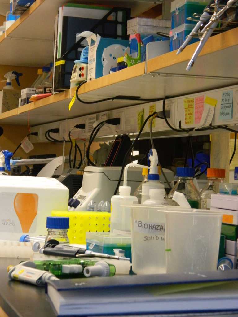
The lights never flicker in the sterile basement hallway. To the left, keypad-guarded doors lead to smaller corridors lined with doors whose double-plated glass windows have been tinted aggressively red. Through those doors, the rooms are lined floor-to-ceiling with mice.
Much like a college dorm, each mouse shares a cage with a few assigned roommates, all of whom are on the same meal plan. Each unit is meticulously labeled and interior decorations are standard. Every single one has a pierced ear. Closer inspection of the silver earrings reveals a truth that sounds as if it came from a science fiction horror blockbuster: these seemingly harmless furballs have been genetically modified.
They could have incredible musical skills or the ability to consume cheese at twice the normal rate! Or they could be ideal models for studying the effects of specific genetic mutations on brain function in the context of human diseases.
Dr. William Dauer, physician, professor, and researcher at the University of Michigan (U of M) is much more interested in the latter. In his neuroscience lab at U of M’s Biomedical Sciences Research building, Dauer and a team of dedicated students and collaborators are using mouse models of human diseases to research movement disorders, which are neurological conditions that affect the higher-level coordination of movement.
“The lab is split into two movement disorders, Parkinson’s Disease and primary dystonia. We’re doing a variety of studies trying to discover how those diseases come about. Our main strategy is to use genes that we know to be mutated in humans and cause those disorders and take those in the lab and try to figure out what they do in the cell,” said Dauer. This is where the mice come in. “We can introduce the mutation that occurs in the patient into the mouse … so we create mice that have the same gene as a human patient and we can study that mouse.”
Mouse brain cells can also be extracted, grown in tubes, and then subjected to additional tests.
“We introduce [a gene] to cells and … look at what other proteins it could interact with,” Dauer said. “We measure things about the cells’ function and ask questions like does the cell die? Does it curl up?”
The tests and questions for the two movement disorders are as different as the diseases themselves, that is, vastly but with many understated similarities.
Parkinson’s disease primarily affects the elderly and in neurodegenerative, meaning that its many symptoms are the result of brain cell death. Primary dystonia, on the other hand, is most commonly found in children between the ages of eight and fourteen and is not thought to be primarily neurodegenerative. It has by definition a single symptom: dystonia, which is a prolonged, involuntary twisting movement of muscles. Dauer, however, finds research on the two to be highly related.
Their most obvious link is that dystonia itself can be a symptom of Parkinson’s disease. Findings related to dystonia could be relevant to understanding and treating that symptom of Parkinson’s. Additionally, research on both diseases adds to the understanding of the life span of individual neurons and the entire brain.
During the initial formation of a human brain, new neurons are created by cell division. Those infant cells must then migrate to their correct location within the brain and begin forming and pruning connections with other neurons in order to create a fully functioning brain.
“Then there are changes at the other end of life, when you’re aging,” Dauer said. “At each of these different stages, neurons are challenged in different ways. There are certain vulnerabilities when a neuron is migrating and others when it is aging. When a neuron is exposed to damage via some pathways it is protected from others.”
Pathways vulnerable during neuron migration are not highly relevant to Parkinson’s patients while processes of aging do not affect primary dystonia. Considered as different parts of a single lifestyle, however, observations of the two diseases could provide insight into the changes and function of the brain.
The brain is arguably the most mystifying and alluring organ in the human body. It is essential to every function of the body yet it is hardly understood. Neurons function and malfunction in fantastic and frustrating ways. At a time in which one out of every eight American develops Alzheimer’s disease yet its ultimate cause remains unknown, neuroscience research is of the utmost importance.
Standing in front of a lab mouse cage and staring into the eyes of a potential data point is a poignant reminder of the humanity in science. Labs like Dr. Dauer’s are in a worldwide relay race against time that is set in a labyrinth and time has already won. We keep going because every mouse could lead to the next breakthrough. There is no hope of ever getting out of the labyrinth but making a single right turn is victory enough to justify the struggle.